K.A. Lidke Lab

K.A. Lidke Laboratory

Optical and analysis methods for measuring the organization, dynamics, and interactions of proteins in living cells.

Research Highlights

These highlights are a selection of the lab's technology development; for our other collaborative work and applications to biology, see the full publications page →.

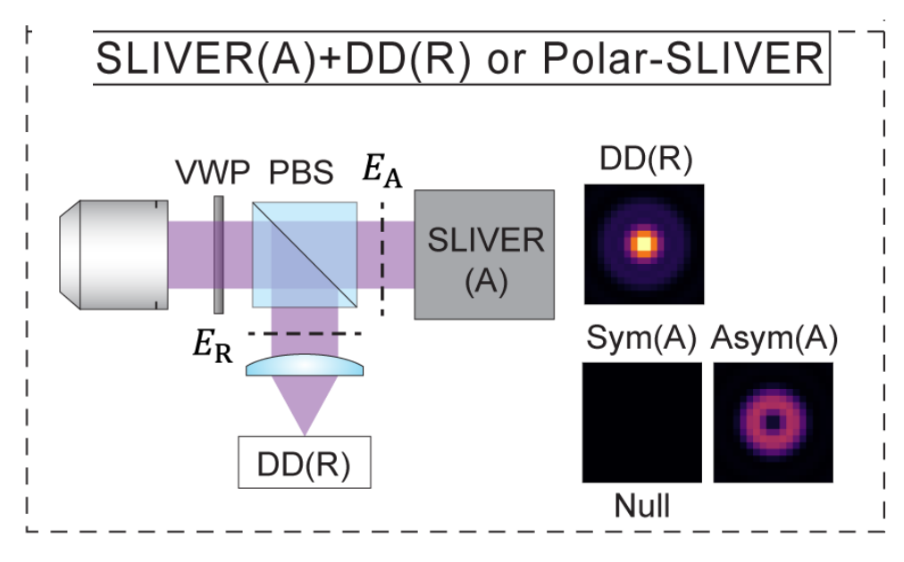
The proposed POLAR-SLIVER measurement system: a vortex wave plate and polarizing beam splitter split the collected light into its azimuthal and radial polarization components; the azimuthal component passes through a SLIVER image-inversion interferometer that sorts it into a symmetric image (which nulls to darkness) and an antisymmetric ring encoding the emitter separation, while the radial component is directly detected (DD). This azimuthal/radial splitting keeps separation-estimation precision near the quantum limit at separations where direct detection alone collapses
2025

Imaging near the quantum limit

In 2016, quantum estimation theory upended Rayleigh's criterion: Tsang, Nair, and co-workers showed that “Rayleigh's curse,” the collapse of separation-estimation precision below the diffraction limit, is not fundamental, and that mode-sorting measurements such as SPADE and SLIVER can reach the quantum Cramér–Rao lower bound (QCRLB), the precision limit for any measurement quantum mechanics allows. But that theory, and its experimental demonstrations, assumed idealized scalar point sources, while the single fluorophores of super-resolution microscopy are freely rotating dipole emitters imaged through high-NA objectives, where the quantum limit was unknown. In Liu et al. (Physical Review A, 2026) we computed the QCRLB for two freely rotating dipoles using vectorial diffraction theory and found, contrary to scalar-model predictions, that standard SLIVER loses its advantage at high numerical aperture because dipole radiation mixes even and odd fields, degrading interference visibility. We introduced Polar-SLIVER, which uses a vortex wave plate to send the purely antisymmetric azimuthal polarization into the inversion interferometer, restoring non-divergent, near quantum-limited precision at any separation, and quantified the effects of background, detection bandwidth, and misalignment. Earlier, in Schodt et al. (Optics Express, 2023), we showed that an image inversion interferometer keeps its precision advantage over direct imaging across a wide range of aberrations and misalignments when pixelated detection is used.
2023–2025

Open-source software for single-molecule imaging and instrument control

The lab develops and maintains open-source software for single-molecule imaging and instrument control. Two of its MATLAB packages have been peer-reviewed in the Journal of Open Source Software: SMITE, which carries single-molecule data from raw frames through localization, drift correction, single-particle tracking, and analysis; and MIC, an object-oriented package for controlling the cameras, stages, and lasers of custom-built microscopes. The lab also maintains the Julia SMLM ecosystem, the MicroscopeControl.jl instrument stack, and Bayesian tools such as BaGoL. A complete list is on the software page.

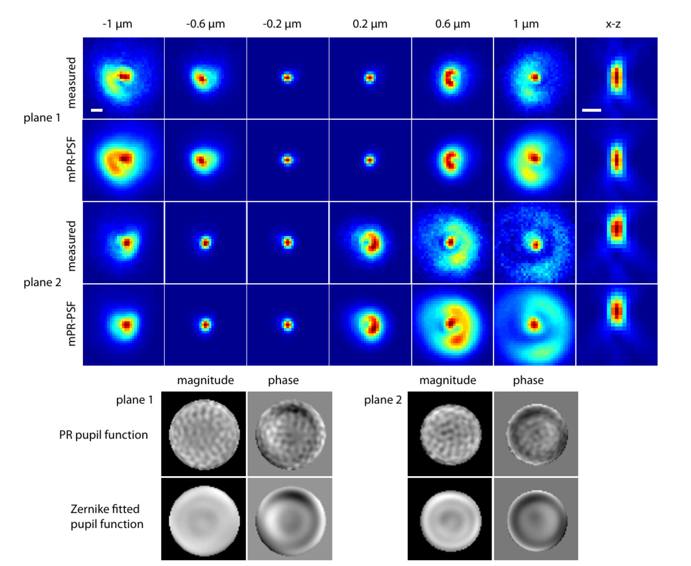
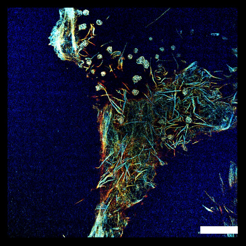
Measured point-spread functions compared with the model phase-retrieved PSF (mPR-PSF) through focus for both focal planes of a biplane microscope, including x-z sections, together with the corresponding phase-retrieved and Zernike-fitted pupil functions (magnitude and phase), showing that a PSF modeled from the instrument's own retrieved pupil reproduces the real, aberrated optics A depth-color-coded three-dimensional super-resolution reconstruction from single-channel supercritical-angle localization microscopy, with axial position running from blue near the coverslip through cyan and yellow to red
2013–2025

Point-spread-function models that match the real microscope

Real optics are never ideal: aberrations distort a microscope's point-spread function (PSF), and they vary across the field of view, so a localization computed from an idealized PSF inherits those errors. We began by replacing idealized models with a PSF retrieved directly from the instrument's own pupil function, which sharpened three-dimensional localization. That work has since grown into a method that infers an accurate PSF directly from the single-molecule data and generalizes across a wide range of microscopes. Most recently, it extends to calibration-free estimation of how aberrations vary across fields of view up to 180 µm, built on nodal aberration theory and a fully vectorial PSF with no bead calibration. The same physics lets us model supercritical-angle fluorescence, the light that molecules within about 100 nm of the coverslip emit into otherwise forbidden angles, so that a single detection channel recovers the axial information that supercritical-angle localization normally needs a two-channel split to obtain.

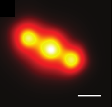
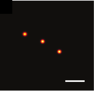
A standard super-resolution reconstruction of a DNA ruler, where three closely spaced docking sites blur together into a single unresolved smear The BaGoL MAPN reconstruction of the same ruler, resolving the three docking sites as individual sharp points
2022

Sub-nanometer precision by Bayesian grouping of localizations

In single-molecule localization microscopy, one emitter blinks and is localized many times, each localization carrying its own error, and a standard reconstruction renders these repeats as a spread of points rather than the single position they came from. BaGoL (Bayesian Grouping of Localizations) treats the repeated blinks of one emitter as noisy measurements of a single position and, with a reversible-jump Markov chain Monte Carlo model, infers how many distinct emitters are present and where each sits, weighting every localization by its own precision. With many repeated, high-precision localizations per emitter, it can infer emitter positions with sub-nanometer precision.

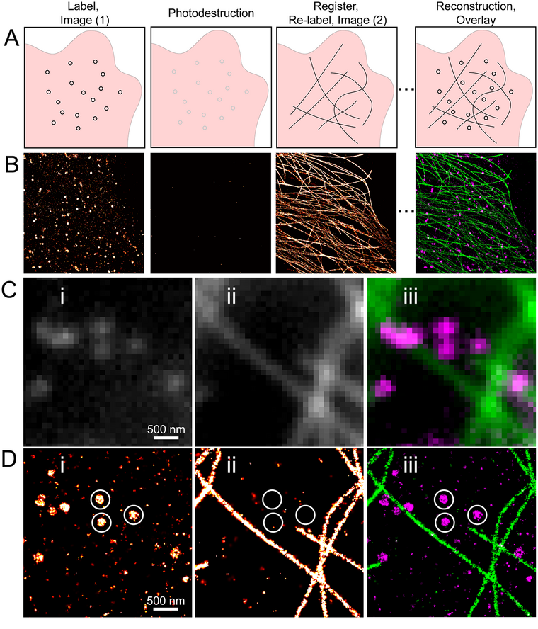
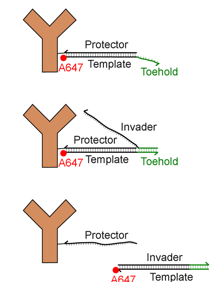
The sequential super-resolution cycle: label and image one target, photo-destroy the label, re-label and image the next, then register and overlay the reconstructions The DNA strand-displacement erase chemistry: an invader strand binds a toehold on the label's template and displaces the protector, gently stripping the dye between rounds
2015–2022

Sequential super-resolution imaging with a single fluorophore

Multi-color super-resolution faces two problems that a single dye avoids. First, photoswitching dyes differ widely in duty cycle, photon yield, bleaching lifetime, and buffer sensitivity, so multiplexing means accepting several imperfect dyes instead of the single best one. Second, and more fundamentally, different colors focus through different optical paths, so multi-color imaging carries chromatic aberration that misregisters the targets at the nanometer scale, a problem inherent to any multi-color readout, including multi-color DNA-PAINT. Sequential super-resolution sidesteps both by imaging every target in turn with the same optimal dye, Alexa Fluor 647, through one optical path, leaving no chromatic aberration between targets and overlaying the rounds to about 10 nm by brightfield correlation. Pallikkuth et al. then replaced the harsh photobleach-and-quench erase with toehold-mediated DNA strand displacement, a fast, gentle, reagent-free way to strip each label between rounds. Schodt et al. built a dedicated microscope that runs the full multi-round acquisition unattended, stabilizing the stage to nanometers and masking regions where the sample itself has moved between rounds.

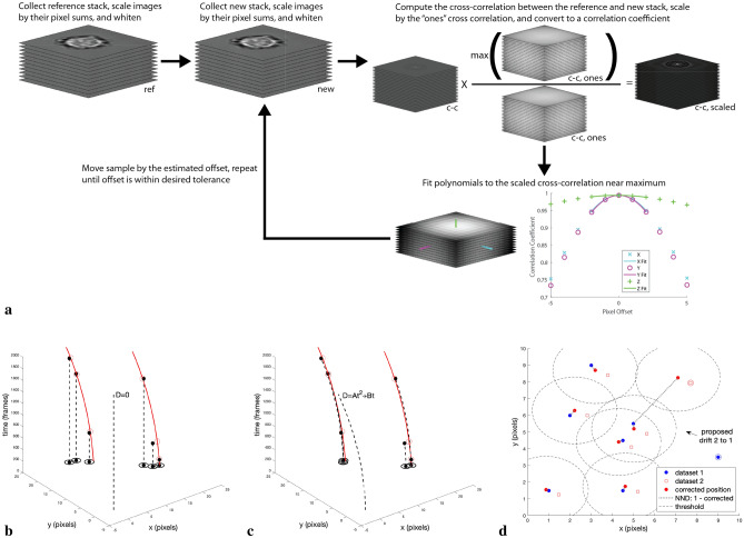
Fiducial-free drift-correction algorithm registering a super-resolution dataset to itself by iterative cross-correlation
2021

Analysis methods for single-molecule localization microscopy

The lab develops analysis methods that turn raw localizations into accurate super-resolution reconstructions. A single blinking molecule is localized many times across consecutive frames; our frame-connection method recognizes these repeats and merges them by solving a linear assignment problem that weights each localization by its precision and by the local emitter density. Sample drift over a long acquisition can exceed the resolution of the reconstruction, so our fiducial-free drift correction registers the localization data to itself, needs no fiducial beads, stays robust even at low localization counts, and gives stable reconstructions over long acquisitions.

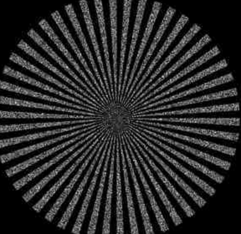
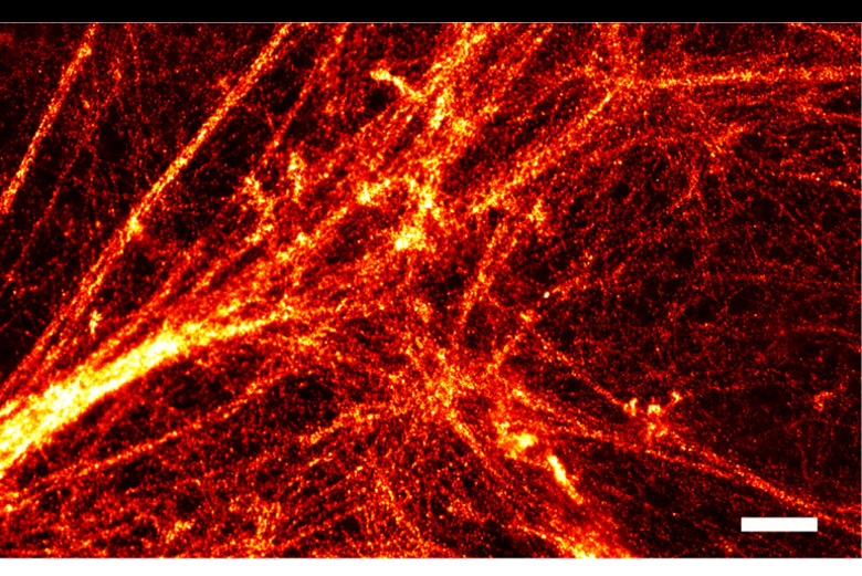
A Siemens-star target with densely packed emitters resolved by multi-emitter fitting A Bayesian multi-emitter (BAMF) super-resolution reconstruction of the actin cytoskeleton
2011–2019

Fitting overlapping emitters at high density

Single-molecule super-resolution is fastest when many molecules are on at once, but then their images overlap, and fitting them one at a time fails. We developed maximum-likelihood fitting that resolves several overlapping emitters within a single region simultaneously, choosing the number of emitters by model selection and raising the tolerable density roughly tenfold. The idea later became fully Bayesian: reversible-jump Markov chain Monte Carlo (BAMF) treats the emitter count itself as unknown, returning a posterior over how many molecules are present and where, together with structured background. In the paper's benchmarks on both simulated and experimental high-density data, BAMF outperformed the established methods FALCON, SRRF, and single-emitter fitting in both detection (Jaccard index) and localization accuracy, with the largest gains for closely spaced emitters and structured background.

Single-objective light sheet formed by a 45-degree mirrored sidewall in a sample channel, folding the illumination sheet into the focal plane of one objective
2016

Single-objective light-sheet microscopy for high-speed whole-cell 3D super-resolution

Light-sheet microscopy gives clean optical sectioning and low photobleaching by illuminating only the focal plane, but it normally needs a second objective crowding the sample. We built a single-objective light sheet: 45° mirrored sidewalls micro-fabricated into a sample channel fold an illumination sheet into the focal plane of one high-NA objective. Cells confined in the channels sit within the sheet with much-reduced out-of-focus background, enabling high-speed whole-cell 3D imaging and improved single-molecule localization, all on an otherwise conventional microscope.

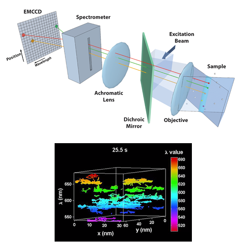
Hyperspectral line-scanning microscope data showing multiple spectrally distinct quantum-dot species separated by emission color
2013

Multi-color quantum-dot tracking using a high-speed hyperspectral line-scanning microscope

Spectral identity is a second axis of independence: probes that overlap in space can still be told apart when their colors differ. We built a high-speed hyperspectral microscope that records a full emission spectrum at every point along a scanned line, and used it to track up to eight spectrally distinct quantum-dot species at once, at 30 frames per second and ~10 nm precision, even where the probes sit closer than the diffraction limit. Because color rather than sparsity does the separating, the time axis stays free for dynamics, so the instrument can follow the assembly and stoichiometry of membrane signaling complexes in live cells with a combination of length scale, speed, and molecular density that no other live-cell method reaches.

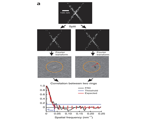
Fourier Ring Correlation curve used to determine the effective resolution of a single-molecule super-resolution reconstruction
2013

Measuring image resolution in optical nanoscopy

A super-resolution image has no single resolution set by the instrument: what you actually resolve depends on labeling density, localization precision, and the structure itself, and for years the field reported it inconsistently. With the Quantitative Imaging group at TU Delft, we adapted Fourier Ring Correlation, long used in cryo-electron microscopy, to single-molecule data: split the localizations into two independent reconstructions and measure the spatial frequency out to which they still agree. The result is a single, sample-specific resolution estimate computed directly from the data, and it has become a standard way to report and compare super-resolution image quality.

Two-color single-quantum-dot trajectories of ErbB1 receptors capturing a dimerization event on a living cell
2011

ErbB1 dimerization is promoted by domain co-confinement and stabilized by ligand

How a growth-factor receptor pair forms and holds together on a living cell had been inferred from structure and bulk measurements rather than watched directly. Using two-color single-quantum-dot tracking, we followed individual ErbB1 (EGFR) receptors and captured their dimerization in real time, then built a three-state hidden Markov model to extract the transition rates among free, co-confined, and dimerized states. The trajectories showed that ligand-bound receptors form markedly more stable dimers than unliganded ones, tying ligand occupancy directly to dimer stability, and that actin confinement promotes dimerization by holding potential partners together long enough to bind.

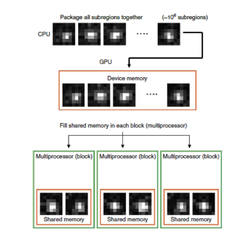
Schematic of GPU parallelization: ~10^6 single-molecule subregions moved into device memory and distributed across multiprocessor blocks with shared memory for simultaneous fitting
2010

Fast, single-molecule localization that achieves theoretically minimum uncertainty

Fitting each single-molecule image by maximum likelihood yields the most accurate position an unbiased estimator can produce (it reaches the Cramér–Rao lower bound, the theoretical minimum uncertainty), but it is costly, so localization software had long fallen back on faster approximations. We showed that the massively parallel architecture of graphics processing units (GPUs) dissolves that trade-off: because every molecule's image is fit independently, thousands can be solved at once. A CUDA implementation, the first to bring GPU computation to single-molecule localization, localized on the order of 105 molecules per second on 2010 hardware while reaching that bound; implementations on current GPUs exceed 106 fits per second. The most demanding step of super-resolution analysis became routine, and with it went any reason to trade accuracy for speed.

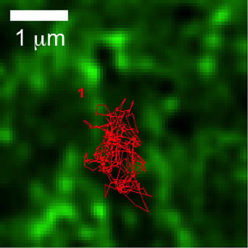
A single quantum-dot-labeled FcεRI receptor trajectory (red) confined within the GFP-labeled cortical actin meshwork (green) in a live mast cell; 1 micron scale bar
2008

Actin restricts FcεRI diffusion and facilitates antigen-induced receptor immobilization

The idea that the cortical actin cytoskeleton confines membrane proteins to transient “corrals” had stood for a decade as inference more than direct observation. By tracking quantum-dot–labeled FcεRI receptors and GFP-tagged actin simultaneously in live mast cells, we caught the two together: actin filaments form dynamic barriers that reorganize over seconds and micrometers, and receptor diffusion changes as those barriers move, direct evidence that actin structures restrict membrane-protein motion in real time. A companion assay measured how fast receptors are immobilized when multivalent antigen cross-links them, and found the response unfolds within seconds and requires an intact actin cytoskeleton. Together the results recast actin from a passive fence into an active participant in receptor diffusion, clustering, and the earliest steps of signaling.

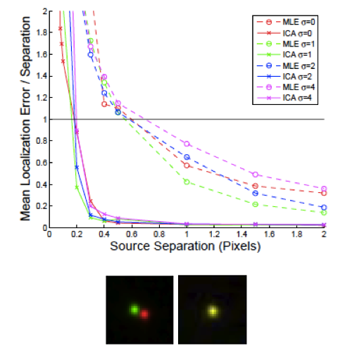
Mean localization error relative to source separation for MLE and ICA methods, with two quantum-dot images resolved below the diffraction limit
2005

Super-resolution by localization of quantum dots using blinking statistics

We were the first to show that the blinking of fluorophores can separate emitters spaced closer than the diffraction limit, localizing each far more accurately than fitting the summed image allows. The method applies independent component analysis (ICA) to an image time series, exploiting the statistically independent intensity fluctuations of neighboring emitters to unmix their overlapping signals, then reconstructing an image from the many recovered positions, an approach we named Pointillism. The demonstration used quantum dots (bright and photostable, but with a high on-fraction that limits usable labeling density), and we proposed that triplet-state blinking and photochromic switching could supply the same independent fluctuations in other labels. The deeper point is one of principle: all resolution is independence. Ordinary imaging separates two points when they are far enough apart to be spatially independent; super-resolution recovers them when they are not, by supplying that independence another way, in this case through the emitters' independent blinking (independence is fundamental; sparsity and stochastic or targeted switching are only means to it).